
Page 42 - Virtual Vascular Vol 17
P. 42
Answers
Figure 3: metallic heart valve (mitral valve) and sternotomy wire
Figure 4: left middle zone mass
Figure 5A: Bilateral bronchopulmonary pneumonia, spinal implant for
previous orthopedic operation.
Nasogastric tube below diaphragm.
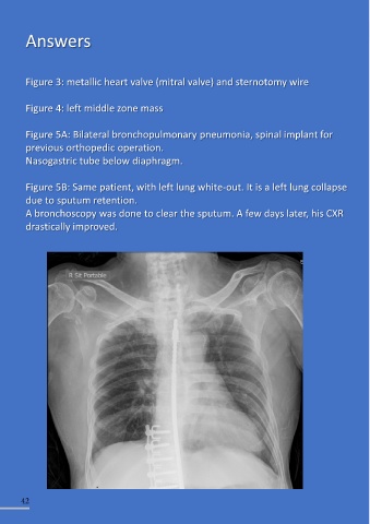
Figure 5B: Same patient, with left lung white-out. It is a left lung collapse
due to sputum retention.
A bronchoscopy was done to clear the sputum. A few days later, his CXR
drastically improved.
42