
Page 41 - Virtual Vascular Vol 6
P. 41
IMAGES IMAGES
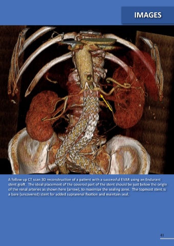
A follow up CT scan 3D reconstruction of a patient with a successful EVAR using an Endurant
stent graft. The ideal placement of the covered part of the stent should be just below the origin
of the renal arteries as shown here (arrow), to maximize the sealing zone. The topmost stent is
a bare (uncovered) stent for added suprarenal fixation and maintain seal.
A follow up CT scan 3D reconstruction of a patient with a successful EVAR showing a
good proximal landing zone (neck), graft landing below the renal arteries, good iliac
landing zone just above (and preserving) both internal iliac artery origin. The calcified
AAA sac is also visualized on this CT.
40 41