
Page 32 - Virtual Vascular Vol 3
P. 32
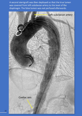
A second stentgraft was then deployed so that the true lumen                                                        Completion angiogram showed that the coeliac axis (CA), superior
               was covered from left subclavian artery to the level of the                                                         mesenteric artery (SMA), right renal (RR) and left renal (LR) arteries

               diaphragm. The false lumen was not perfused afterwards.                                                             were all perfused.


                                                                       Left subclavian artery


                                                                                                                                                                                                                splenic artery
                                                                                                                                                                                            CA                  from CA


                                                                                                                                          RRA
                                                                                                                                                                                                               LRA


                                                                                                                                                      SMA


                            Coeliac axis


        32                                                                                                                                                                                                                            33