
Page 67 - Virtual Vascular Vol 6
P. 67
In this instance, the centre of the base is yellow in colour, and has some slough sounded
with granulation tissue. Students should be able to describe the ulcer in terms of size, site,
edge, base, surrounding tissue, and also and offer differential diagnoses.
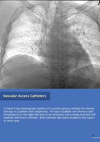
Vascular Access Catheters
A Chest X-ray showing two options of a central venous catheter for chemo-
therapy in a patient with lymphoma. He had a Cephalic vein Porta-a-cath
(Powerport) on the right side due to be removed, and a newly inserted Left
cephalic Hickman’s catheter. Both catheter tips were located in the superi-
or vena cava.
66 67