
Page 15 - Virtual Vascular Volume 7
P. 15
Percutaneous EVAR Aortic Anatomy
L renal vein
IMA
PERCUTANEOUS EVAR became the norm with modern small profile devices.
This photo showed two small groin puncture wounds in a patient who re-
ceived a percutaneous EVAR.
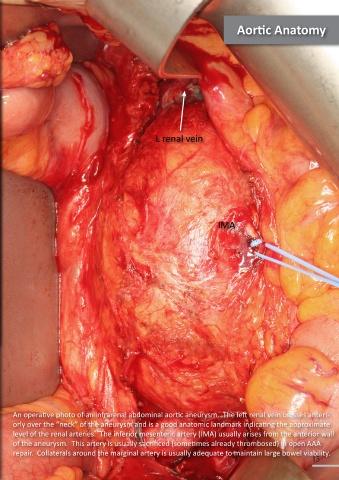
An operative photo of an infrarenal abdominal aortic aneurysm. The left renal vein crosses anteri-
orly over the “neck” of the aneurysm and is a good anatomic landmark indicating the approximate
level of the renal arteries. The inferior mesenteric artery (IMA) usually arises from the anterior wall
of the aneurysm. This artery is usually sacrificed (sometimes already thrombosed) in open AAA
repair. Collaterals around the marginal artery is usually adequate to maintain large bowel viability.
14 15