
Page 35 - Virtual Vascular Volume 7
P. 35
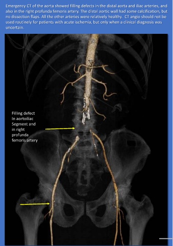
Emergency CT of the aorta showed filling defects in the distal aorta and iliac arteries, and

     also in the right profunda femoris artery. The distal aortic wall had some calcification, but

 Acute Lower Limb Ischemia  no dissection flaps. All the other arteries were relatively healthy.  CT angio should not be
     used routinely for patients with acute ischemia, but only when a clinical diagnosis was
     uncertain.


 An elderly patient presented with a few hours of acute bilateral lower limb
 ischaemia with pain, pallor, paresthesia, and coldness. He denied any history of
 palpitations or angina, backpain or abdominal discomfort, or previous claudication,

 but was hypertensive. His femoral pulses were not palpable, and his feet were cold
 and pale.


         Filling defect
         In aortoiliac

         Segment and
         in right
         profunda

         femoris artery


 What are the differential diagnoses?


 34                                                                                                            35