Page 22 - Virtual Vascular Vol 8
P. 22

Pulmonary Embolism


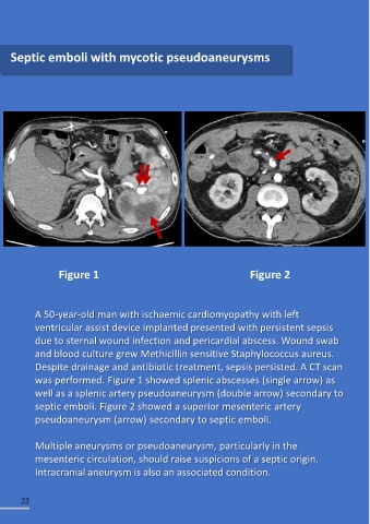
    Septic emboli with mycotic pseudoaneurysms
                                                                                                                                                                                          A patient was admitted with

                                                                                                                                                                                          extensive deep vein thrombosis

                                                                                                                                                                                          involving the left lower limb. He
                                                                                                                                                                                          also presented with severe

                                                                                                                                                                                          shortness of breath and chest
                                                                                                                                                                                          pain associated with sinus

                                                                                                                                                                                          tachycardia


                                                                                                                                                 Figure 1


                                                                                                                                                                                          This is the CT scan finding: Serial
                                                                                                                                                                                          scan from Figure 1 to Figure 3

                                                                                                                                                                                          show massive pulmonary

                                                                                                                                                                                          embolism involving the right
                                                                                                                                                                                          pulmonary artery (arrow)


                    Figure 1                                                     Figure 2


                                                                                                                                                                                          Apart from systemic

            A 50-year-old man with ischaemic cardiomyopathy with left                                                                                                                     anticoagulation, in
            ventricular assist device implanted presented with persistent sepsis                                                                                                          haemodynamically unstable

            due to sternal wound infection and pericardial abscess. Wound swab                                                                  Figure 2                                  patients with massive pulmonary

            and blood culture grew Methicillin sensitive Staphylococcus aureus.                                                                                                           embolism, active  intervention
            Despite drainage and antibiotic treatment, sepsis persisted. A CT scan                                                                                                        with open pulmonary

            was performed. Figure 1 showed splenic abscesses (single arrow) as                                                                                                            embolectomy, systemic
            well as a splenic artery pseudoaneurysm (double arrow) secondary to                                                                                                           thrombolysis or catheter-

            septic emboli. Figure 2 showed a superior mesenteric artery                                                                                                                   directed thrombolysis/

            pseudoaneurysm (arrow) secondary to septic emboli.                                                                                                                            thrombectomy should also be
                                                                                                                                                                                          considered

            Multiple aneurysms or pseudoaneurysm, particularly in the

            mesenteric circulation, should raise suspicions of a septic origin.
            Intracranial aneurysm is also an associated condition.


        22                                                                                                                                                                                                                            23

                                                                                                                                                 Figure 3
   17   18   19   20   21   22   23   24   25   26   27