
Page 24 - Virtual Vascular Vol 8
P. 24
IMAGES
A plain X-ray of this thigh showed also cal-
cifications in the wall of his superficial and
deep femoral arteries (above).
A CT lower limb angiogram revealed the
lower limb arterial anatomy outlined by
calcium. This investigation is expensive
and incurred radiation exposure. The cal-
cium can cause significant artifacts and ac-
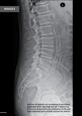
A 66-year old diabetic man complaining of intermittent curate lumen assessment is not possible.
claudication of this right thigh and calf. A lateral X-ray This is the limitation of lower limb CTA
of the spine showed extensive calcifications on the wall which should not be performed routinely
24 of the abdominal aorta and both common iliac arteries. for diagnostic purposes. 25