
Page 11 - Virtual Vascular Vol 15
P. 11
2012 2014
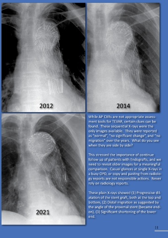
While AP CXRs are not appropriate assess-
ment tools for TEVAR, certain clues can be
found. These sequential X-rays were the
only images available. They were reported
as “normal”, “no significant change”, and “no
migration” over the years. What do you see
when they are side by side?
This stressed the importance of continue
follow up of patients with Endografts, and we
need to revisit older images for a meaningful
comparison. Casual glances at single X-rays in
a busy OPD, or copy and pasting from radiolo-
gy reports are not responsible actions. Never
rely on radiology reports.
These plain X-rays showed (1) Progressive dil-
atation of the stent graft, both at the top and
bottom, (2) Distal migration as suggested by
the angle of the proximal stent (became end-
2021 on), (3) Significant shortening of the lower
end.
11