
Page 29 - Virtual Vascular Vol 15
P. 29
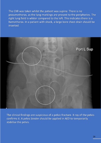
The CXR was taken whilst the patient was supine. There is no
           pneumothorax, as the lung markings are present to the peripheries. The

           right lung field is whiter compared to the left. This indicates there is a

           hemothorax. In a patient with shock, a large bore chest drain should be
           inserted.


         The clinical findings are suspicious of a pelvic fracture. X-ray of the pelvis

         confirms it. A pelvic binder should be applied in AED to temporarily
         stabilise the pelvis.


                                                                                                               29