
Page 33 - Virtual Vascular Vol 16
P. 33
v
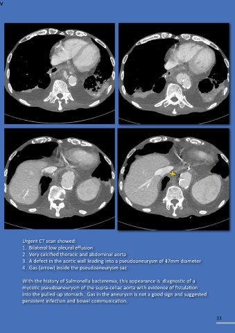
Urgent CT scan showed:
1. Bilateral low pleural effusion
2. Very calcified thoracic and abdominal aorta
3. A defect in the aortic wall leading into a pseudoaneurysm of 47mm diameter
4. Gas (arrow) inside the pseudoaneurysm sac
With the history of Salmonella bacteremia, this appearance is diagnostic of a
mycotic pseudoaneurysm of the supra-celiac aorta with evidence of fistulation
into the pulled-up stomach. Gas in the aneurysm is not a good sign and suggested
persistent infection and bowel communication.
33