
Page 40 - Virtual Vascular Vol 16
P. 40
Pulmonary Embolism
Figure 1
Figure 2
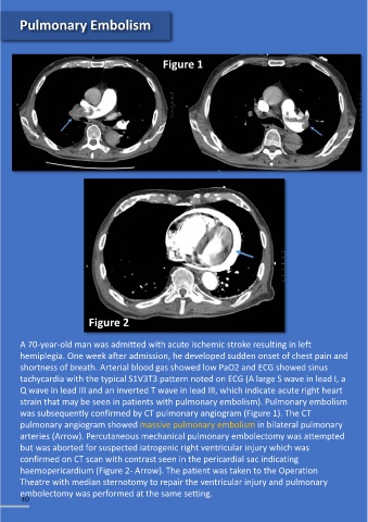
A 70-year-old man was admitted with acute ischemic stroke resulting in left
hemiplegia. One week after admission, he developed sudden onset of chest pain and
shortness of breath. Arterial blood gas showed low PaO2 and ECG showed sinus
tachycardia with the typical S1V3T3 pattern noted on ECG (A large S wave in lead I, a
Q wave in lead III and an inverted T wave in lead III, which indicate acute right heart
strain that may be seen in patients with pulmonary embolism). Pulmonary embolism
was subsequently confirmed by CT pulmonary angiogram (Figure 1). The CT
pulmonary angiogram showed massive pulmonary embolism in bilateral pulmonary
arteries (Arrow). Percutaneous mechanical pulmonary embolectomy was attempted
but was aborted for suspected iatrogenic right ventricular injury which was
confirmed on CT scan with contrast seen in the pericardial sac indicating
haemopericardium (Figure 2- Arrow). The patient was taken to the Operation
Theatre with median sternotomy to repair the ventricular injury and pulmonary
embolectomy was performed at the same setting.
40