
Page 71 - Virtual Vascular Vol 16
P. 71
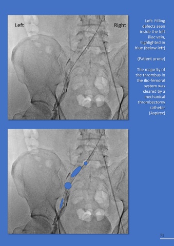
This venogram (left) shows                                                                         Left: Filling
 the IVC filter after   Left                                                   Right              defects seen
 deployment (via a right                                                                         inside the left

 femoral vein approach).                                                                              iliac vein,
                                                                                                 highlighted in
                                                                                             blue (below left)


                                                                                               (Patient prone)
 The angiogram (below left)

 shows multiple filling                                                                        The majority of
 defects in the left femoral                                                                  the thrombus in

 vein highlighted in blue                                                                      the ilio-femoral
 (below right).  The                                                                                system was
 venogram was via left                                                                             cleared by a
 popliteal vein USS guided                                                                          mechanical

 puncture (Prone position)                                                                     thrombectomy
                                                                                                        catheter
                                                                                                       (Aspirex)


                                                                                                               71