Page 67 - Virtual Vascular Vol 16
P. 67

On further history taking, he had no weight loss or symptoms suspicious of
             malignancy.  There was no abdominal mass and per-rectal exam was also
             normal.


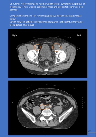
             Compare the right and left femoral and iliac veins in the CT scan images
             below.
             Notice how the left side is hypodense compared to the right, signifying a

             filling defect (thrombus).


                Right                                                                            Left


                                                                                                               67
   62   63   64   65   66   67   68   69   70   71   72