Page 70 - Virtual Vascular Vol 16
P. 70

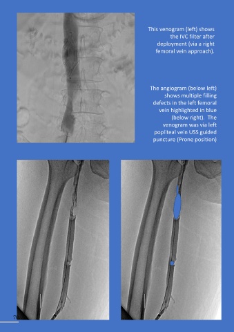
This venogram (left) shows
                                                                                        the IVC filter after
                                                                                 deployment (via a right

                                                                                femoral vein approach).


                                                                             The angiogram (below left)

                                                                                     shows multiple filling
                                                                               defects in the left femoral

                                                                                  vein highlighted in blue
                                                                                        (below right).  The
                                                                                    venogram was via left
                                                                               popliteal vein USS guided

                                                                               puncture (Prone position)


        70
   65   66   67   68   69   70   71   72   73   74   75