
Page 75 - Virtual Vascular Vol 11
P. 75
3
2
1
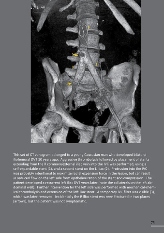
This set of CT venogram belonged to a young Caucasian man who developed bilateral
iliofemoral DVT 10 years ago. Aggressive thrombolysis followed by placement of stents
extending from the R common/external iliac vein into the IVC was performed, using a
self-expandable stent (1), and a second stent on the L iliac (2). Protrusion into the IVC
was probably intentional to maximize radial expansion force in the lesion, but can result
in reduced flow on the left side from epitheliorization of the stent and compression. The
patient developed a recurrent left iliac DVT years later (note the collaterals on the left ab-
dominal wall). Further intervention for the left side was performed with mechanical-chem-
ical thrombolysis and extension of the left iliac stent. A temporary IVC filter was visible (3),
which was later removed. Incidentally the R iliac stent was seen fractured in two places
(arrows), but the patient was not symptomatic.
75