
Page 73 - Virtual Vascular Vol 11
P. 73
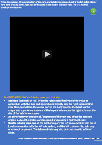
Three-dimensional CT reconstruction of the aorta and inferior vena cava, showing the left-sided inferior
  vena cava, crossing to the right side of the aorta at the level of the renal vein. This is  a normal
  developmental variant.


             IVC2.mp4


      MALFORMATIONS of the inferior vena cava include:

      •    Agenesis (absence) of IVC- when the right subcardinal vein fail to make its
           connection with the liver and shunts blood directly into the right supracardinal
           vein. Thus, blood from the caudal part of the body reaches the heart via the

           azygos and superior vena cava and the hepatic vein enters the right atrium at the
           site of the inferior vena cava
      •    An abnormality of position of / segments of the vein may affect the adjacent

           organs, such as the ureter, compressing it and causing a hydronephrosis
      •    Double inferior vena cava at the lumbar region: the left sacro-cardinal vein fail to
           lose its connection with the left subcardinal, and the left common iliac vein may
           or may not be present. The left renal vein may also be in retro-aortic in 5% of

           cases.


                                                                                                               73
                      Pansky B. Review of medical embryology. Chapter 126. Development of The Venous System: The Inferior Vena Cava