
Page 20 - Virtual Vascular Vol 15
P. 20
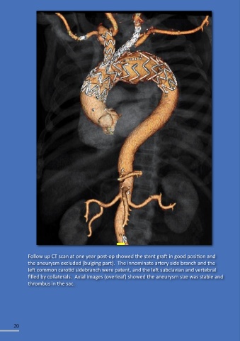
Follow up CT scan at one year post-op showed the stent graft in good position and
               the aneurysm excluded (bulging part).  The innominate artery side branch and the
               left common carotid sidebranch were patent, and the left subclavian and vertebral
               filled by collaterals.  Axial images (overleaf) showed the aneurysm size was stable and
               thrombus in the sac.


        20