
Page 23 - PowerPoint Presentation
P. 23
Liposarcoma
Illustration
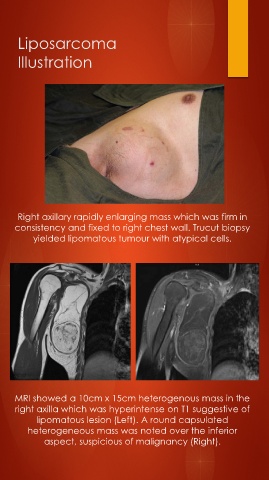
Right axillary rapidly enlarging mass which was firm in
consistency and fixed to right chest wall. Trucut biopsy
yielded lipomatous tumour with atypical cells.
MRI showed a 10cm x 15cm heterogenous mass in the
right axilla which was hyperintense on T1 suggestive of
lipomatous lesion (Left). A round capsulated
heterogeneous mass was noted over the inferior
aspect, suspicious of malignancy (Right).