
Page 14 - PowerPoint Presentation
P. 14
Desmoid tumour
Illustration
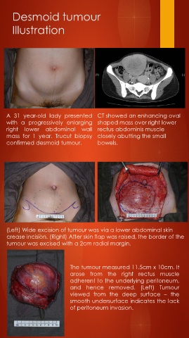
A 31 year-old lady presented CT showed an enhancing oval
with a progressively enlarging shaped mass over right lower
right lower abdominal wall rectus abdominis muscle
mass for 1 year. Trucut biopsy closely abutting the small
confirmed desmoid tumour. bowels.
(Left) Wide excision of tumour was via a lower abdominal skin
crease incision. (Right) After skin flap was raised, the border of the
tumour was excised with a 2cm radial margin.
The tumour measured 11.5cm x 10cm. It
arose from the right rectus muscle
adherent to the underlying peritoneum,
and hence removed. (Left) Tumour
viewed from the deep surface – the
smooth undersurface indicates the lack
of peritoneum invasion.